New ultrasound capability brings research and welfare benefits to preclinical imaging
A new ultrasound imaging system is expanding the University of Southampton’s preclinical research capabilities, giving researchers new ways to study disease and experimental treatments while also supporting improvements in animal welfare.
Funded through a Medical Research Council grant led by Professor Nick Evans, the S-Sharp Prospect T2 is based within the Biomedical Research Facility (BRF) and supported by the Biomedical Imaging Unit (BIU). Both are part of Southampton Medical Research Services.
Designed specifically for high-resolution imaging in small animals, the system enables researchers to examine mice and rats in real time. It can produce both 2D and 3D images, while Doppler and contrast-enhanced ultrasound can be used to investigate blood flow and perfusion, and elastography can measure tissue stiffness.
The system adds ultrasound to Southampton’s existing preclinical X-ray CT and optical imaging technologies, allowing researchers to combine complementary approaches to build a more complete picture of the biological processes they are investigating.
This opens up potential applications across cancer, musculoskeletal and regenerative medicine, liver disease, cardiovascular medicine, neuroscience and ophthalmology.
Discussing this award, Professor Evans said: “This is an important addition to Southampton’s research infrastructure, giving researchers across a wide range of disciplines access to a powerful new imaging capability. It will help us study disease and evaluate new treatments in greater detail, while supporting high-quality, responsible research.”
Following change over time
One of the most important benefits of the new system is its ability to take repeated measurements from the same animal, allowing researchers to follow biological changes over time.
In some studies, collecting tissue requires a terminal procedure, meaning the same animal cannot be studied again at a later stage. Researchers may therefore need to use different animals to understand how disease or treatment develops over time.
Ultrasound can provide an alternative by allowing researchers to follow some of these changes in the same living animal. A cancer researcher, for example, could identify a tumour, measure its volume and then monitor how it changes during an experimental treatment.
Researchers could similarly investigate blood flow changes, track how tissue becomes stiffer as disease develops, or examine how heart function changes over the course of an experiment.
Collecting this kind of longitudinal data can provide a more complete picture of disease progression and treatment response. At the same time, where repeated imaging can replace the need for separate animals at different time points, it creates opportunities to reduce the number of animals needed for some studies and to refine the way experiments are carried out.
Dr Katie Dexter, who manages the system day to day, says its value therefore goes beyond the images and research data it can generate.
“It’s not just good for research and data, it’s good for welfare as well.”
These benefits support the 3Rs principles of replacement, reduction and refinement, which underpin responsible animal research.
Improving precision and developing skills
The benefits of being able to see inside the body in real time also extend to experimental procedures.
The equipment supports ultrasound-guided injections, where a needle is aligned with the ultrasound probe so that researchers can see both the needle and its intended target on screen. This allows cells, drugs or other agents to be delivered more precisely to specific locations.
The same capability can also support training. Researchers learning injection techniques can receive visual feedback on whether they have reached the correct location, helping them develop their skills before carrying out procedures independently.
Greater accuracy and consistency in these procedures has the potential to benefit both the quality of the resulting research and animal welfare.
Taking the technology to the research
To make these capabilities useful across Southampton’s preclinical research, the system has also been designed with portability in mind.
Research takes place across different areas of the Biomedical Research Facility, with strict biosecurity controls governing the movement of animals between environments. Rather than unnecessarily moving animals to imaging equipment, the ultrasound system can be taken closer to where the research is happening.
Built around a tablet unit, it can be moved between appropriate areas of the BRF following the necessary cleaning and decontamination procedures. Separate supporting equipment has also been purchased for different animal areas to minimise what needs to be transferred.
This flexibility means researchers can make use of a single imaging system across different areas of the facility while maintaining the controls required for animal research.
Supporting welfare alongside research
Alongside its experimental applications, ultrasound can help with welfare monitoring, including pregnancy assessment and the investigation of potential health concerns. Depending on the procedure, imaging may also be possible without prolonged anaesthesia.
Where anaesthesia is required, the system incorporates ECG and respiration monitoring, while its platform can be heated to help maintain the animal’s temperature. The animal also remains visible and accessible to trained staff throughout imaging.
Together, these capabilities give researchers and technical staff more options to monitor animals, refine procedures and generate high-quality scientific results.
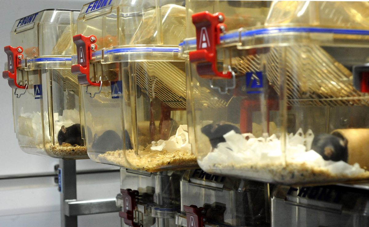
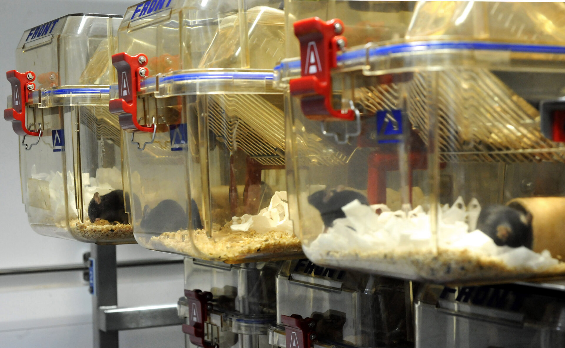
Laboratory mice in cages. Image courtesy of Understanding Animal Research, licensed under Creative Commons Attribution 4.0.